We look back at some of 2025’s major advances in AI-driven live cell analytics, from automated mitochondrial segmentation to dynamic EC50 profiling and next-generation T-cell insights. These breakthroughs bring deeper, label-free understanding to drug discovery and immuno-oncology.
AI accelerated the analysis of live cell images
in 2025
More groups adopted Nanolive’s AI-powered solutions to analyze their label-free live cell images in 2025, and 25% of these peer-reviewed papers included quantification of cellular phenotypic features from Nanolive images. Automated analysis by Nanolive’s digital assays was used to study mitochondrial metabolism, cell phenotypes in disease models, and lipid metabolic shift, amongst other applications.
Author Dai-Wei Hu, at the China Medical University published a study into early-onset breast cancer in Advanced Science, and shared the group’s opinion on the benefits of using Nanolive’s AI, the impact it had on user workflow, and data reproducibility with us:
“The AI-based mitochondrial analysis software significantly accelerated our data analysis workflow. The automated quantification of mitochondrial parameters enabled us to rapidly obtain objective and reproducible quantitative metrics, reducing manual bias and increasing the overall interpretability and reliability of the results. […] Overall, we found that Nanolive served as a robust and user-friendly platform”. – Dai-Wei Hu
To learn more about Nanolive’s digital analysis solutions for cellular and organelle phenotyping, visit https://v11pzd3gpyc.c.updraftclone.com/technology/solutions-for-biotech-and-pharma/.
To read the full paper, click here (open access).
AI image analysis reinforces molecular methods for greater biological relevance and data confidence
One growing trend in 2025 that is expected to continue into 2026 was the integration of AI image analysis alongside other molecular biology methods such as transcriptomics, metabolomics, and cell painting to increase biological relevance and robustness of data interpretation. Imaging cells at high resolution can help to verify whether molecular signatures observed in omics-based screens translate into measurable phenotypic changes, or serve as an initial phenotypic reference to guide downstream pathway analysis..
A group at the Broad Institute of MIT published the paper “Integrated spatial morpho-transcriptomics predicts functional traits in pancreatic cancer” which utilized Nanolive imaging, fluorescent cell painting, and transcriptomics as complementary methods to perform phenotypic profiling of different cell lines and treatment conditions. The authors wrote: “In conclusion, our SMART approach highlights the exciting possibility of leveraging integrated morphologic, transcriptional, and functional measurements on multiple patient avatars in response to drug treatment to comprehensively model patient disease in real-time.”
You can read the full text published in Science Advances here (open access).
Nanolive imaging can be used to profile cell phenotypes at the organelle, cell, and population levels, and to cross-compare phenotypic metrics across biological dimensions (for example mitochondrial state, lipid organization, and overall cell health), including through integrated analyses. To download a 1-page guide to label-free cell painting, click here: Form: Label-free phenotyping angle one pager

Label-free live cell imaging as a reference method
in 2025’s research
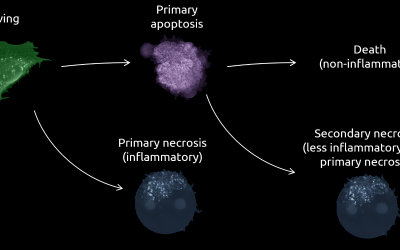
Label-free live cell imaging continued to be widely adopted approach for studying cell phenotypes and dynamics in 2025, with over 100 new publications added by Nanolive’s clients, reaching over 460 in total. Among the reported applications enabled by Nanolive’s advanced imaging and analysis technologies, mitochondria, metabolism, and immuno-oncology research were the most popular fields, highlighting the recognition of noninvasive imaging as a key tool for studying mitochondria and immune cells where fluorescent labeling can alter cellular behavior. Investigations into cell death and drug discovery, also frequently used noninvasive timelapse imaging to monitor dynamic cellular responses, supporting the assessment of cell death mechanisms, drug effects, and phenotype stability over time.

In addition, Nanolive’s technology also gained recognition across the pharmaceutical and academic sectors, with established companies and institutes such as Amgen and MIT contributing to this year’s publication record in high-impact journals including Nature, Advanced Science, Theranostics, Nature Communications, and the Journal for Immunotherapy of Cancer.
The following scheme summarizes the key words that featured the most in publications this year:

Latest publication highlights with Nanolive imaging:
- Phenotypic profiling: Gong, D. et al. (2025) ‘Integrated spatial morpho-transcriptomics predicts functional traits in pancreatic cancer’, Science Advances, https://doi.org/10.1126/sciadv.adx0632
- Metabolic reprogramming: Yang, C. et al. (2025) ‘Pulsed electromagnetic fields modulate energy metabolism during wound healing process: an in vitro model study’, Scientific Reportshttps://doi.org/10.1038/s41598-024-69862-x
- Immunotherapy: Yang, C. et al. (2025) ‘A trispecific antibody engaging T cells with tumour and myeloid cells augments antitumour immunity’ Nature Biomedical Engineering, https://doi.org/10.1038/s41551-025-01569-4
- Drug mechanism of action: Michel, O. et al. (2025) ‘Application of nisin as a potential drug candidate for electrochemotherapy’ Frontiers in Oncology, https://doi.org/10.3389/fonc.2025.1689261
- Drug delivery: Klebowski, B. et al. (2026) ‘Effect of the porosity of the palladium shell in Au-core Pd-shell bimetallic nanoparticles for the radiosensitizing properties in simulated anticancer proton radiotherapy’ Colloids and Surfaces B: Biointerfaces, https://doi.org/10.1016/j.colsurfb.2025.115192
Find over 400 publications featuring Nanolive imaging here.
Subscribe to this monthly newsletter to stay up to date with AI applications in cell biology by clicking the button at the bottom of the page.
Read our latest news
Cell Health and Stress Application Note
A cutting-edge approach to cell health and stress with Nanolive’s LIVE Cytotoxicity Assay Discover the advanced capabilities of Nanolive’s LIVE Cytotoxicity Assay in our latest application note. This document presents a detailed exploration of how our innovative,...
Investigative Toxicology Application Note
Introducing the LIVE Cytotoxicity Assay: A breakthrough in investigative toxicology Discover the forefront of drug safety and toxicology evaluation with Nanolive's LIVE Cytotoxicity Assay Application Note. Our groundbreaking approach offers a label-free, high-content...