Microglia-neuron interactions play a key role in brain health and disease
Understanding microglial function is a research priority for neuroscientists (1, 2). Microglia-neuron interactions play a key role in keeping the brain healthy, and modifications in microglial morphology are often signs of neurodegenerative disease.
Nanolive label-free live cell imaging reveals dynamic microglia-neuron interactions in real time
It is particularly difficult to perform ectopic gene expression within microglia primarily because they are very difficult to transfect, which has to date hampered the use of live cell imaging to study their interaction with neurons in real time. Nanolive imaging avoids this limitation because it is label-free. The result is beautiful footage like this video, shot during a demonstration with the Hanus and the Van Niel labs at the Institute of Psychiatry and Neurosciences in Paris.
The video reveals the dynamic interactions between human microglial 3 (HMG3) cells and 3-week-old rat hippocampal neurons. One image was taken every 30 secs for 10 h using our 3D Cell Explorer-fluo.
Microglia play a key-role in modulating neuron activity
HMG3 are the dense, bright cells sitting on top of the neurons. HMG3 are highly motile and constantly scan the neural network for molecular cues indicative of homeostatic disturbance with outstretched filopodia (3). Such behavior is coherent with their biological function as the resident immune cells of the brain. These macrophage-like cells play a key role in modulating neuron activity, trimming synaptic elements (e.g. dendritic spines), phagocytosing dying cells and helping to coordinate the inflammation response (4).
Microglia are long-lived cells that self-renew (4). In the video, we observe one HMG3 cell undergoing mitosis (cell bottom right, second from the right). At second 10 the cell nucleus rotates, at second 11 the cell undergoes prophase and at second 13 the chromosomes line up along the metaphase plate.
The ability to observe neuron-microglia interactions label-free over long time frames opens the door to understanding how defects in microglia function could contribute to or trigger diseases (3).
References
(1) Pósfai, B., Cserép, C., Orsolits, B., & Dénes, Á. (2019). New insights into microglia–neuron interactions: a neuron’s perspective. Neuroscience, 405, 103-117.
(2) Südhof, T. C. (2017). Molecular neuroscience in the 21st century: a personal perspective. Neuron, 96(3), 536-541.
(3) Bernier, L. P., Bohlen, C. J., York, E. M., Choi, H. B., Kamyabi, A., Dissing-Olesen, L., Hefendehl, J. K., Collins, H. Y., Stevens, B., Barres, B. A., MacVicar, B. A. (2019). Nanoscale surveillance of the brain by microglia via cAMP-regulated filopodia. Cell Reports, 27(10), 2895-2908.
(4) Szepesi, Z., Manouchehrian, O., Bachiller, S., & Deierborg, T. (2018). Bidirectional microglia–neuron communication in health and disease. Frontiers in Cellular Neuroscience, 12, 323.
Read our latest news
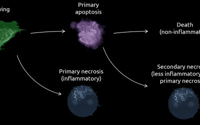
Cell Health and Stress Application Note
A cutting-edge approach to cell health and stress with Nanolive’s LIVE Cytotoxicity Assay Discover the advanced capabilities of Nanolive’s LIVE Cytotoxicity Assay in our latest application note. This document presents a detailed exploration of how our innovative,...
Investigative Toxicology Application Note
Introducing the LIVE Cytotoxicity Assay: A breakthrough in investigative toxicology Discover the forefront of drug safety and toxicology evaluation with Nanolive's LIVE Cytotoxicity Assay Application Note. Our groundbreaking approach offers a label-free, high-content...

Trends in AI analysis for live cell imaging
Nanolive microscopes
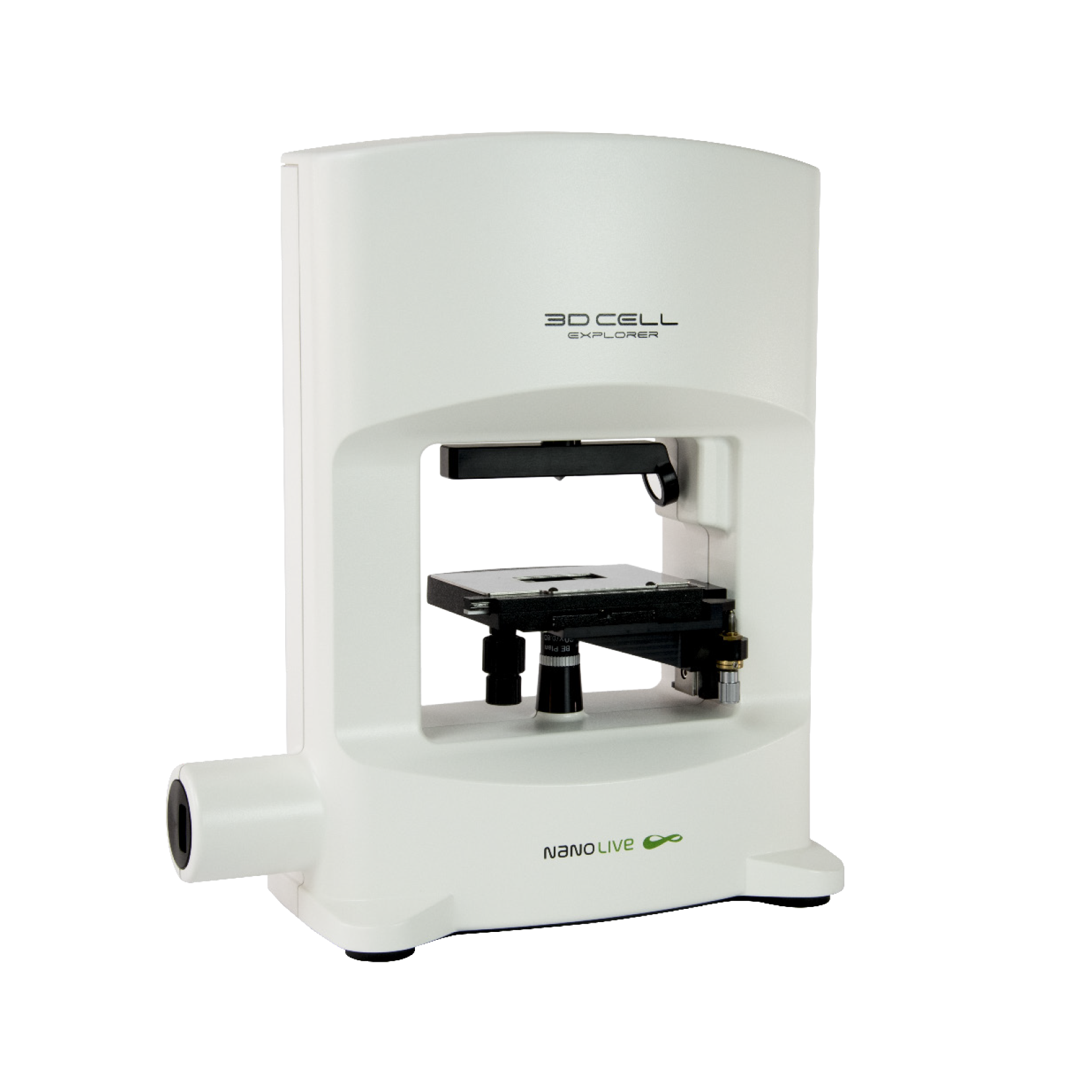
3D CELL EXPLORER
Budget-friendly, easy-to-use, compact solution for high quality non-invasive 4D live cell imaging
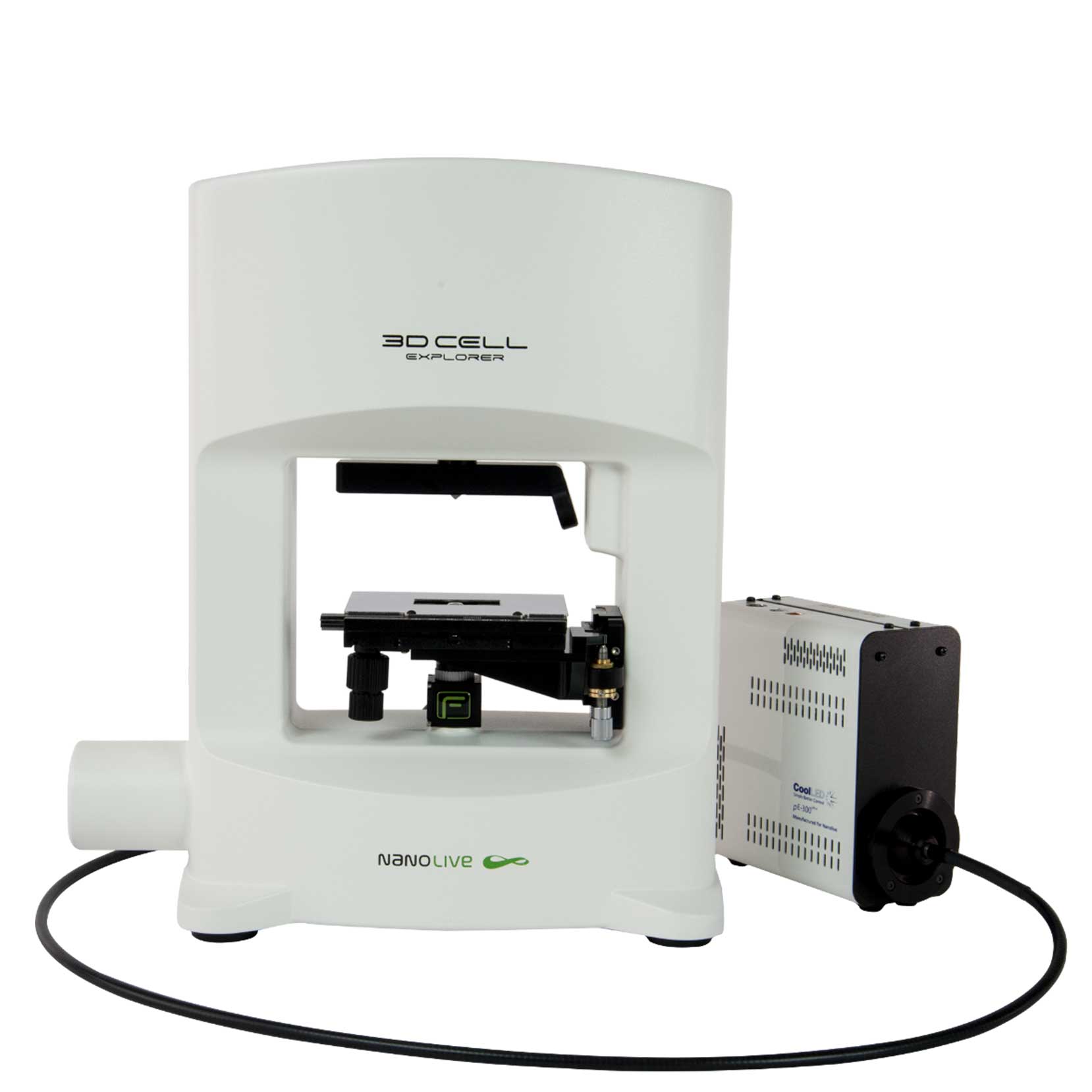
3D CELL EXPLORER-fluo
Multimodal Complete Solution: combine high quality non-invasive 4D live cell imaging with fluorescence
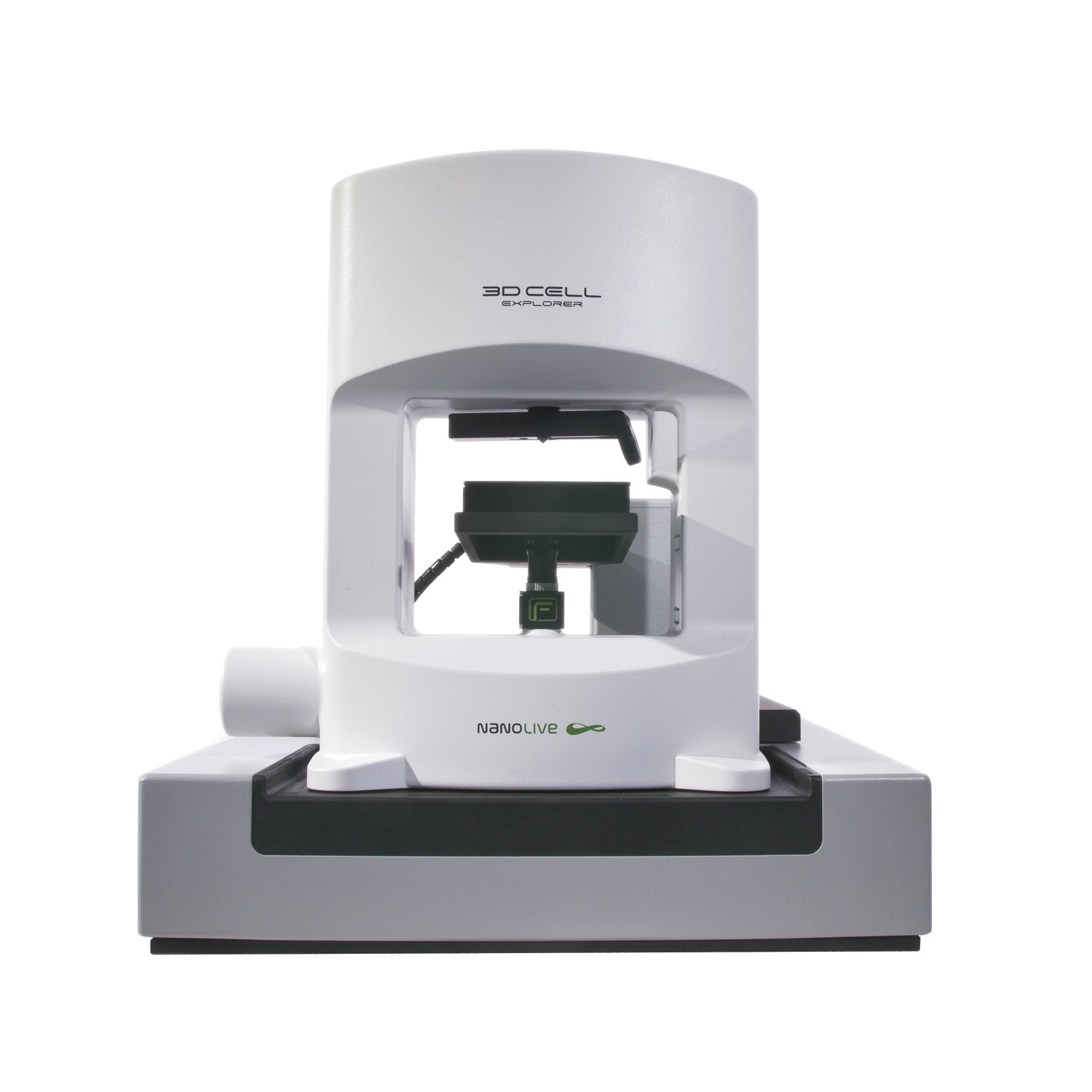
CX-A
Automated live cell imaging: a unique walk-away solution for long-term live cell imaging of single cells and cell populations